ENVIRONMENTAL CHAMBER TECHNICAL FEATURES
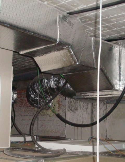
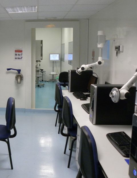
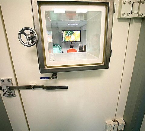
The Controlled Environment Research Laboratory (CER-Lab) of Vision R&D, SL. located at Edificio IOBA, Campus Miguel Delibes, Paseo Belén 17, 47011 Valladolid, Spain, is able to create, control and monitor typical indoor and outdoor environmental conditions. The CER-Lab is composed of the Exposure Room where Temperature (T), Relative Humidity (RH), Air Flow, Illuminance and Pressure is controlled, and the Assessment Room, where T and RH are kept similar to those of the Exposure Room.

Exposure room
- Temperature (T). Range: from 15⁰C to 30⁰C. One ⁰C steps.
- Relative Humidity (RH). Range: from 5% to 80%. One % steps.
- Air flow. Range: Lack – turbulent air flow.
- Illuminance. Range: from 50 to 1000 lux. One lux steps.
- Pressure. Range: from 940 mbar (750 m above sea level) to 450mbar (6500m above sea level). Ability to reach 750 mbar (pressure inside a conventional airplane cabin). One mbar steps.
Evaluation room
- Temperature (T). Range: from 15⁰C to 30⁰C. One ⁰C steps.
- Relative Humidity (RH). Range: from 5% to 60%. One % steps.
- Long-duration exposure studies. Tº and RH are maintained.

- High precision analytical balance & antivibration table
- Internal calibration
- Readability and repeatability of 0.1mg
- Stabilization time 3.5 sc
- Belmonte noncontact gas esthesiometer.
- Wider temperature range: 0-99.9ºC
- Wider mechanical range
- Confocal in vivo scanning laser microscopy (HRT-III) with Rostock cornea module (Heidelberg).
- The instrument can be converted into a confocal corneal microscope using an additional microscope lens which attaches to the standard lens.
- Along with corneal analysis software, the HRT is able to image cells and cell layers within the cornea.
- Osmometers (Tearlab and Tear Osmometer 3100).
- TearLab has a positive predictive value of 89%.
- The advanced tear Osmometer 3100 has a resolution of 1 mOsm/kg H2O with a linearity less than 2% from a straight line and a Standard Deviation less than or equal to 5 mOsm/kg H2O (Range: 280 to 350 mOsm/kg H2O)
- Tearscope (Keeler).
- Hemispherical cup of 90mm
- Handle with a central 15mm diameter observation hole
- Sample obtaining
- Microcaps for tears
- Polyethersulfone filters and Cytobrush Plus for conjunctival cells
Within the exposure room, up to 8 subjects can perform different visual tasks (playing cards, reading, watching TV, etc) under tightly controlled conditions; within the assessment room, patients are evaluated prior to and after exposure using the following Diagnostic Instruments:
- Digital LogMAR ETDRS chart (Topcon CC-100XP):
- Visual Acuity test: Letters, Tumbling E, Landolt C and Numbers. Masking is available for horizontal (row), vertical (column) and single characters.
- Contrast Sensitivity test: Spatial Frequency Contrast Sensitivity Test can be customized by setting the amount of spatial frequencies and contras levels.
- Computerized corneal topographer (Medmont E300):
- Coverage Diameter: 0.25 – 11 mm
- Field of View: 11mm H x 11mm V
- Power Range: 10-100 Diopters
- Number of Rings: 32
- Measurement Points: 15,120
- Working Distance: 30mm
- Repeatability: Test Object < 0.1 Diopters
- Hartmann-Shack wavefront aberrometer (irx3, Imagine eyes):
- 1024 true optical measurement points.
- Wide dynamic range of ±10D astigmatism and -16/+20D sphere.
- Repeatability of 0.003D RMS in the laboratory.
- Video slit lamp system (Topcon SL-8z):
- Digital imaging system
- Magnification knob ergonomically positioned
- Magnification changes of 6.35X to 31.75X.
- Filter choices include: cobalt blue, redfree, 13% neutral density, and a heat absorption filter
- Expanded viewing field
- 316mm working distance


ANCILLARY LABORATORIES AND UNITS
- Ocular Immunology Clinical & Contact Lens Unit
It is a twelve full-time team members group (Ocular Surface Group) coordinated by two ophthalmologists, who are able to perform advanced diagnostic, therapeutic and surgical procedures. In addition, they lead clinical trials and supervise immune-related laboratory studies.
- Molecular Biology Lab
It is composed of three specialized team members who provide laboratory support to the Ocular Immunology Clinical Unit. They are able to perform Flow Cytometry, real time PCR, ELISA and Immunofluorescence techniques.
- Eye Pathology Lab
It is managed by an expertly trained team dedicated exclusively to the evaluation of ocular tissue.
- Clinical Trials Unit
It is composed of three high qualified research members. It was certified by the European Vision Institute. Clinical Trials. Sites of Excellence (EVI.CT.SE.) Network and has already carried out 27 trials since 2005.

